
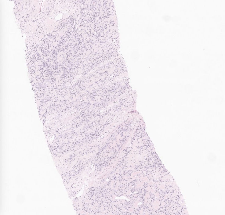
A 30-year-old female with an incidental finding of supraclavicular mass with calcification.


What's your morphological impression at this point?
A: Nodular fasciitis
B: Atypical fibroxanthoma
C: Low grade fibromyxoid neoplasm
D: Low grade fibromyxoid sarcoma
E: Myxoma
F: Solitary fibrous tumor
G: Ossifying fibromyxoid tumor
H: Angiofibroma of soft tissue
I: Myxoid diffuse neurofibroma
Answer
An extended IHC panel showed the tumors are positive for CD10 only,
while negative for CD34, STAT6, SOX10, S100, MUC4, SMA, myogenin,
beta catenin (nuclear staining), p63, and pankeratin. RB1 is retained. The
tumor cells are focal and weakly positive for ER and desmin.
What's the diagnosis?
A: Nodular fasciitis
B: Atypical fibroxanthoma
C: Low grade fibromyxoid neoplasm
D: Low grade fibromyxoid sarcoma
E: Myxoma
F: Solitary fibrous tumor
G: Ossifying fibromyxoid tumor
H: Angiofibroma of soft tissue
I: Myxoid diffuse neurofibroma
Answer
Case was sent to sarcoma fusion panel and NBPF10::PHF1 fusion was detected. What are the tumors associated with this fusion?
A: Ossifying fibromyxoid tumor
B: Superficial acral fibromyxoma
C: Chondromyxoid fibroma
D: Intramuscular myxoma
E: Cutaneous nerve sheath myxoma
F: Myxofibrosarcoma
Answer
References:
https://www.pathologyoutlines.com/topic/softtissueossifyingfibromyxoid.html
https://tumourclassification.iarc.who.int/chaptercontent/33/117
Folpe AL, Weiss SW. Ossifying fibromyxoid tumor of soft parts: a clinicopathologic study of 70 cases with emphasis on atypical and malignant variants. Am J Surg Pathol. 2003 Apr;27(4):421-31. doi: 10.1097/00000478-200304000-00001. PMID: 12657926.
Miettinen M, Finnell V, Fetsch JF. Ossifying fibromyxoid tumor of soft parts--a clinicopathologic and immunohistochemical study of 104 cases with long-term follow-up and a critical review of the literature. Am J Surg Pathol. 2008 Jul;32(7):996-1005. doi: 10.1097/PAS.0b013e318160736a. PMID: 18469710.
Case credit: UCSD Pathology
Author: Wangpan Jackson Shi, MD
Comments